At Smiles N Beyond in Wallington, NJ, we prioritize diagnostic clarity because precise images lead to better treatment decisions. Cone-beam computed tomography (CBCT) gives our team three-dimensional views of teeth, jaws, and surrounding structures that traditional two-dimensional X-rays simply cannot provide. Used judiciously, CBCT helps us see relationships and details that matter for planning restorative, surgical, and orthodontic care while keeping patient comfort and safety front and center.
Conventional dental X-rays are excellent for many routine needs, but they compress complex anatomy into a flat image. CBCT captures volumetric data, allowing clinicians to examine anatomy from multiple angles and in true spatial context. That dimensional insight reduces guesswork: clinicians can visualize root canals, bone contours, and sinus anatomy with clarity, which supports more confident diagnoses and fewer surprises during treatment.
This 3D information is particularly valuable when anatomic variations or hidden pathology could affect care. For example, a tooth that appears straightforward on a two-dimensional film may reveal a previously unseen canal or an atypical root formation on a CBCT scan. Early identification of these nuances helps shape a treatment strategy that is precise and conservative.
Importantly, CBCT is a diagnostic tool, not a replacement for clinical judgment. Our team integrates imaging findings with a thorough clinical exam and patient history to form a complete picture before recommending any next steps. When used appropriately, 3D imaging elevates the quality and predictability of care across many dental specialties.
Planning complex procedures—whether placing implants, preparing for oral surgery, or evaluating impacted teeth—benefits from the spatial accuracy CBCT provides. With a volumetric scan, clinicians can measure bone volume and density, locate vital structures such as nerves and the sinus floor, and determine the optimal orientation and depth for surgical or restorative work. That level of detail helps reduce intraoperative adjustments and enhances outcome predictability.
CBCT data can be exported to planning software that simulates procedures and supports the creation of surgical guides. By rehearsing the treatment in a virtual environment, clinicians can anticipate challenges and refine their approach. This digital-first planning often translates into shorter chair time and fewer surprises during treatment.
Beyond surgical planning, CBCT can assist with complex restorative and endodontic decisions. When tooth anatomy is atypical or pathology is suspected, three-dimensional imaging clarifies the extent of involvement and helps determine whether conservative treatment or a more involved approach is necessary. In short, CBCT enables more informed decisions that align with each patient’s unique anatomy.
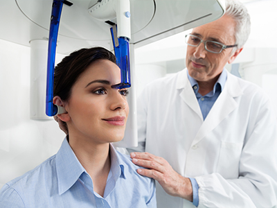
A CBCT scan is a quick and uncomplicated experience for most patients. Unlike full-body CT scanners, dental CBCT units are compact and designed specifically for head and neck imaging. During the scan, you will either sit or stand comfortably while the unit rotates around your head. The entire acquisition typically takes only a minute or less, and there is no need for uncomfortable positioning devices in most cases.
Radiation dose is an important consideration, and modern CBCT systems are engineered to be efficient. They focus on the area of interest and use exposure settings appropriate for dental applications, which helps minimize patient exposure while still producing diagnostic-quality images. Our team follows established safety protocols and applies imaging only when the expected clinical benefit outweighs any risk.
After the scan, the raw data are reconstructed into detailed images that can be reviewed in multiple planes or rendered into three-dimensional models. Because the process is so rapid, the imaging component rarely adds significant time to an appointment, yet it often yields information that substantially improves the diagnostic and planning phases of care.
CBCT produces rich datasets, but valuable information depends on expert interpretation. Reading three-dimensional images requires training and experience to distinguish normal anatomic variation from clinically significant findings. Our clinicians evaluate scans in the context of your symptoms, clinical exam, and dental history to draw conclusions that are relevant to your care.
Interpretation often involves collaboration. Specialists in endodontics, oral surgery, orthodontics, or radiology may be consulted when complex anatomy or pathology is identified. This multidisciplinary approach ensures that imaging findings are translated into practical, patient-centered treatment recommendations rather than isolated observations.
In addition to diagnostic review, CBCT images can be integrated with other digital tools—such as intraoral scans and treatment-planning software—to produce surgical guides, custom restorations, and visual treatment previews. This integration streamlines workflows and improves communication between clinicians and patients about the proposed plan.
CBCT has broad utility across dentistry. In implant dentistry, it helps assess bone volume and angulation, identify ideal implant positions, and avoid critical structures. For endodontic care, CBCT reveals complex canal morphologies, root fractures, and periapical pathology that may not be visible on standard films. Orthodontists use three-dimensional data to evaluate tooth position, skeletal relationships, and airway considerations when planning comprehensive treatment.
Oral and maxillofacial surgeons rely on CBCT for evaluating impacted teeth, planning extractions, and assessing trauma or pathology of the jaws. It is also a tool for evaluating temporomandibular joint (TMJ) anatomy and for airway analysis in cases where breathing-related concerns intersect with dental care. In every case, CBCT adds a level of detail that supports safer, more tailored interventions.
Because each application has unique requirements, our team determines the appropriate field of view and imaging parameters based on the clinical question. This targeted approach ensures that every scan is designed to answer the specific diagnostic need while minimizing unnecessary exposure.
CBCT is a powerful diagnostic resource when applied thoughtfully and interpreted by experienced clinicians. At Smiles N Beyond, our commitment is to use advanced imaging to inform careful, patient-focused care in Wallington, NJ. If you have questions about whether CBCT is appropriate for your situation or would like more information about how we use 3D imaging in treatment planning, please contact us to learn more.


CBCT, or cone-beam computed tomography, is a specialized imaging technology that captures three-dimensional volumetric images of the teeth, jaws and surrounding structures. Unlike conventional two-dimensional dental X-rays, CBCT acquires data from multiple angles and reconstructs it into true spatial views that reveal depth, relationships and internal anatomy. This three-dimensional perspective helps clinicians detect details that can be obscured or flattened on standard films.
Because CBCT shows anatomy in multiple planes, it makes measurements of bone volume, angulation and proximity to vital structures more accurate. That added precision supports diagnostic clarity and reduces uncertainty when planning complex restorative, surgical or endodontic care. CBCT is therefore a complementary diagnostic tool used alongside clinical examination and other imaging modalities.
CBCT is typically recommended when two-dimensional images and a clinical exam do not provide sufficient information to answer a specific diagnostic question. Common indications include implant planning, evaluation of impacted or aberrant teeth, assessment of suspected root fractures or complex canal anatomy, and investigation of jaw pathology. The decision to image is always guided by the clinical problem and the expectation that three-dimensional data will affect the treatment approach.
Clinicians weigh the potential diagnostic benefit against radiation exposure and tailor the field of view and imaging parameters to the clinical need. CBCT is not used for routine exams but is reserved for situations where volumetric detail materially improves decision-making. When appropriate, it enhances safety, predictability and the likelihood of a favorable outcome.
In implant planning, CBCT provides precise information about bone height, width and density as well as the location of nerves and the maxillary sinus. Those measurements allow clinicians to determine optimal implant size, position and angulation while avoiding critical anatomy. This three-dimensional planning reduces the need for intraoperative adjustments and supports a more predictable restorative result.
CBCT datasets can be integrated with planning software to simulate implant placement and design surgical guides when indicated. By rehearsing the procedure virtually, the team can anticipate challenges and communicate a clear plan with laboratory partners and specialists. That digital workflow improves coordination and typically reduces chair time during the actual surgery.
CBCT units designed for dental use deliver substantially lower radiation doses than conventional medical CT scanners, but exposure is still an important consideration that we manage proactively. Clinicians follow the ALARA principle—keeping exposure as low as reasonably achievable—by selecting the smallest field of view and lowest acceptable exposure settings for the diagnostic task. Modern machines also incorporate dose-reduction technologies and patient positioning features that further limit unnecessary exposure.
Before ordering a scan, the care team evaluates whether CBCT will meaningfully influence diagnosis or treatment planning and documents the clinical justification. When imaging is indicated, protocols are customized to the patient and the problem, and scans are limited to the area of interest to avoid excess radiation. This targeted, case-by-case approach balances diagnostic benefit with patient safety.
A CBCT scan is typically quick and straightforward. Patients either sit or stand while the scanner rotates around the head for a brief period, often under one minute for acquisition, and no invasive positioning devices are usually required. The process is noninvasive, and most people experience little or no discomfort during imaging.
After acquisition, the raw data are reconstructed into multiplanar views and three-dimensional renderings that clinicians can review immediately. Because the scan itself is rapid, it rarely adds significant time to an appointment but can provide information that shortens or streamlines subsequent treatment steps. The clinician will discuss the findings and how they relate to the proposed care plan.
Interpreting CBCT studies requires training and experience to distinguish normal anatomic variations from clinically significant findings. At our office, clinicians review scans in the context of the patient’s symptoms, clinical examination and dental history to arrive at meaningful conclusions that inform treatment. When scans reveal complex or ambiguous findings, the team may consult with specialists in endodontics, oral surgery, orthodontics or oral and maxillofacial radiology for additional expertise.
Interpretation often leads directly to treatment recommendations, such as altered surgical approach, referral for specialty care or targeted conservative management. CBCT images are also used as part of a collaborative planning workflow, enabling clear communication among treating clinicians and with the patient. Reports and visualizations are documented in the chart to support continuity of care.
Yes. CBCT can reveal anatomic details and pathology that are difficult or impossible to appreciate on two-dimensional radiographs. Examples include hidden root canals, vertical root fractures, small periapical lesions, complex root morphologies and the spatial relationship of impacted teeth to adjacent structures. These discoveries can change a clinician’s diagnosis or treatment plan and reduce the risk of unforeseen complications.
CBCT is also valuable for evaluating the temporomandibular joint, airway space and bony lesions of the jaws, areas where conventional films can be limited by overlap and lack of depth information. While CBCT is not the best modality for soft-tissue detail in all cases, its strength in depicting bone and dental anatomy often makes it the preferred tool when three-dimensional clarity is required. Clinicians select imaging that best answers the clinical question at hand.
CBCT is excellent for visualizing hard tissues, but it has limitations that clinicians must consider. Soft-tissue contrast is inferior to medical CT or MRI, and metallic restorations can produce artifacts that obscure adjacent anatomy. The effective diagnostic value also depends on correct field-of-view selection, image quality and proper interpretation by trained clinicians.
CBCT should not replace a thorough clinical examination and history; rather, it complements those assessments. In some scenarios, additional imaging such as medical CT or MRI may be necessary to evaluate soft-tissue pathology or complex conditions. Understanding these limitations helps clinicians choose the most appropriate imaging pathway for each patient.
CBCT data can be exported and merged with intraoral scans and other digital records to create a comprehensive three-dimensional model for treatment planning. This integration allows clinicians to visualize both hard and soft tissues together, design restorations, and plan implant positioning with a high degree of precision. When surgical guides are indicated, the combined datasets form the basis for guide fabrication that translates virtual plans into accurate intraoperative execution.
That digital workflow enhances communication between the dental team, laboratories and specialists and supports predictable restorative outcomes. It also permits patient education by showing clear visualizations of anatomy and proposed interventions. Ultimately, the interoperability of CBCT with other digital tools streamlines complex care and improves coordination.
If you have symptoms or a treatment need that may benefit from three-dimensional imaging, schedule a consultation with our team in Wallington, NJ so a clinician can perform a focused exam. During the visit, the clinician will review your dental and medical history, perform an intraoral and extraoral examination, and determine whether CBCT will add information that influences diagnosis or treatment. That clinical evaluation is the first step in deciding whether a scan is appropriate.
When a CBCT scan is recommended, the care team will explain the purpose of the study, what to expect during imaging, and how the results will be used in planning your care. If additional specialist input is indicated, clinicians will coordinate consultations to ensure imaging findings are translated into a clear, patient-centered plan. This approach helps ensure imaging is used thoughtfully and only when it benefits the patient’s care at Smiles N Beyond.

Ready to schedule your next dental appointment or have questions about our services?
Getting in touch with Smiles N Beyond is simple. Our friendly team is here to help you schedule appointments, answer questions about treatment options, and address any concerns you may have. Whether you prefer to call our office, send an email, or complete our easy online contact form, we’re happy to assist you. Take the first step toward a healthier, more confident smile, contact us today and experience the difference personalized dental care can make.