An intraoral camera is a compact, pen-sized imaging tool that captures high-resolution, full-color views of the inside of the mouth. Unlike traditional dental mirrors, this small camera provides magnified visuals that reveal fine details of tooth surfaces, gum tissue, and restorative work. The images appear instantly on a monitor, giving both the clinician and patient a clear, shared view of oral conditions that would otherwise be difficult to inspect at a glance.
Because the device records real-time video and still photos, it transforms what was once an abstract verbal description into concrete visual evidence. This clarity helps clinicians identify early signs of decay, hairline fractures, worn restorations, and areas of inflammation with greater confidence. The result is more precise examinations and a stronger foundation for diagnostic decision-making.
Modern intraoral cameras are designed to be ergonomic, easy to maneuver around the mouth, and gentle on soft tissues. Their portability and high-quality optics make them an integral part of a contemporary dental exam, allowing for fast, noninvasive imaging that complements standard radiographs and clinical evaluation.
One of the most practical benefits of intraoral imaging is how it reshapes patient communication. Instead of relying solely on descriptions or diagrams, clinicians can show patients actual photographs of their teeth and gums. This shared visual language removes ambiguity, helps patients understand the nature and location of concerns, and supports more informed treatment planning.
When patients can see the problem for themselves—whether it’s early decay, a cracked tooth, or gum recession—they are often more comfortable participating in discussions about care options. Visual evidence also helps set realistic expectations about outcomes and timelines, which can reduce anxiety and build trust in the treatment process.
Documentation captured by the camera can also be used to track changes over time. Sequential images taken at different visits create a visual timeline that helps patients appreciate progress after treatment or notice gradual changes that require attention. This continuity strengthens the patient–provider relationship and encourages proactive oral health habits.
Intraoral cameras integrate smoothly with modern dental record systems, allowing stills and video clips to be saved directly into a patient’s chart. These images complement X-rays and 3D scans by providing surface-level detail that radiographs cannot, such as color variations, plaque accumulation, and soft-tissue condition. Together, these tools create a fuller diagnostic picture.
High-quality intraoral images are also useful for interdisciplinary care. When collaboration with a specialist or dental laboratory is necessary, clinicians can share annotated photos to clarify findings and treatment goals. This precise communication can streamline referrals and lab prescriptions by reducing misunderstandings about shade, margin location, or tissue contours.
Because digital capture is immediate and portable, intraoral camera data supports faster clinical workflows. Electronic records populated with clear images enhance documentation accuracy, improve case acceptance conversations, and provide defensible clinical evidence should questions arise during treatment or insurance review.
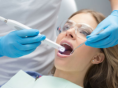
A typical camera-assisted exam is quick and comfortable. The clinician or hygienist will gently position the device inside your mouth and sweep it over the teeth and soft tissues while the images appear on a screen. Most captures take only seconds, and as necessary, the clinician may take additional close-ups of specific areas to record fine details or compare against prior visits.
There is no radiation involved with intraoral photography, and the procedure is noninvasive. Patients may be asked to open slightly or help with angling by turning their head, but overall the process is suited to virtually all ages. Images can be reviewed immediately, and the clinician will explain what each photo shows and why a particular observation is clinically relevant.
Because the images become part of the permanent record, they are available for later review, second opinions, or treatment planning sessions. This transparency lets patients take a more active role in decisions about restorative, preventive, or cosmetic care when those conversations arise.
At Smiles N Beyond, we view intraoral cameras as more than a gadget; they are a clinical standard that supports meticulous care. By combining visual documentation with clinical expertise, we can detect subtle problems sooner and tailor treatments more precisely. The technology reinforces our commitment to clear communication and patient-centered decision-making.
Using intraoral imagery aligns with a philosophy of prevention and minimally invasive dentistry: early detection often means simpler, less invasive treatment. When conditions are identified before they progress, care can focus on preservation and stabilization rather than emergency intervention. This approach benefits patients by protecting long-term oral health and reducing avoidable complications.
Incorporating high-quality visual records into routine exams also helps the team maintain consistent, evidence-based standards across providers and visits. Whether for monitoring restorative margins, assessing tissue health, or documenting healing after treatment, intraoral photos are a reliable, reproducible tool that supports excellent clinical outcomes.
Summary: Intraoral cameras bring a real-time, magnified view of the mouth to the dental exam, improving diagnostics, documentation, and patient communication. By combining these clear images with clinical insight, your dental team can identify problems earlier, explain treatment options more effectively, and monitor progress over time. If you’d like to learn more about how intraoral imaging is used in our office, please contact us for more information.


An intraoral camera is a small, pen-sized imaging device that captures high-resolution, full-color views of the inside of the mouth. It provides magnified, real-time stills and video that appear instantly on a chairside monitor. These images reveal fine details of tooth surfaces, restorations, and soft tissues that are difficult to see with traditional mirrors.
Most systems combine LED illumination with a compact digital sensor and ergonomic optics to produce clear images in tight spaces. Clinicians can freeze frames, take close-ups, and record video to document findings and support diagnostic decisions. The portability and ease of use make intraoral cameras a routine part of modern dental examinations.
Intraoral imaging helps detect early signs of decay, hairline fractures, worn restorations, and areas of inflammation before problems progress. By providing a visual record, the technology reduces guesswork and increases diagnostic confidence. Patients and clinicians benefit from a shared view that supports clearer communication and more accurate treatment planning.
Captured images also serve as a baseline for monitoring changes over time and evaluating treatment outcomes. They can streamline referrals and lab communication by showing exact margin locations and tissue contours. Overall, intraoral cameras contribute to more conservative, evidence-based care.
When patients can see high-resolution photos of their own teeth and gums, complex findings become easier to understand. Visual evidence removes ambiguity that often accompanies verbal descriptions and diagrams. That shared perspective encourages informed questions and more meaningful discussions about care options.
At Smiles N Beyond, clinicians review images chairside to explain conditions, illustrate proposed treatments, and set realistic expectations. Annotated photos and side-by-side comparisons help patients weigh alternatives and consent to plans with confidence. This transparent approach strengthens trust and supports long-term oral health decisions.
Intraoral imaging is noninvasive and involves no ionizing radiation because it records only surface-level photos and video. Most individual captures take only a few seconds and are well tolerated by patients of all ages. The small, ergonomic cameras are designed to be gentle on soft tissues and easy to position in the mouth.
Clinics follow strict infection-control protocols, using disposable sheaths or sterilized barriers for each use to maintain hygiene. Staff are trained to operate the device efficiently to minimize discomfort and exam time. If a patient is sensitive or anxious, the clinician can pause, explain each step, and proceed at a comfortable pace.
Intraoral photos capture color, surface texture, plaque, and soft-tissue details that radiographs and 3D scans cannot show. X-rays reveal internal structures such as root anatomy and bone levels, while intraoral images document external conditions and visual cues. Together they provide a comprehensive diagnostic picture that supports precise treatment planning.
For example, photos are useful for shade matching, evaluating margins, and documenting tissue health before and after procedures. When clinicians combine these surface images with radiographs or CBCT scans, they can correlate visual findings with internal anatomy to make more informed decisions. This multimodal approach reduces uncertainty and improves clinical outcomes.
During a camera-assisted exam, a hygienist or dentist will gently position the device and sweep it across teeth and soft tissues while images appear on a monitor. Most individual captures take only a few seconds, and clinicians may take additional close-ups of areas of concern. You may be asked to slightly adjust your head or open wider to improve angles, but the process is generally quick and comfortable.
The clinician will review key images with you immediately, pointing out findings and explaining why they matter for treatment or prevention. Selected photos are saved to your chart for future comparison or referral purposes. This immediate feedback helps patients participate in care decisions and track progress over time.
Most modern practices integrate intraoral photos directly into the electronic dental record, where stills and video clips are linked to specific exams and treatment notes. These images become part of your permanent chart and are available for later review during follow-up visits. Properly labeled images improve documentation accuracy and continuity of care across providers.
Clinicians can annotate images, compare them side-by-side with prior visits, and include them when coordinating care with specialists or dental laboratories. Secure systems allow controlled sharing for consultation while maintaining patient privacy and record integrity. This organized visual documentation supports clearer treatment plans and more reliable monitoring of oral health changes.
By revealing early surface changes like demineralization, hairline cracks, or localized plaque, intraoral cameras enable intervention before problems require extensive treatment. Early detection often allows for conservative treatments such as sealants, targeted fluoride, or minimally invasive restorations. Photographic records also reinforce oral hygiene instruction by showing patients exactly where to focus their home care.
In follow-up visits, images document healing and treatment effectiveness, allowing clinicians to adjust care plans without unnecessary delays. This proactive documentation supports a philosophy of preservation and long-term tissue health. The result is often fewer invasive procedures and better outcomes for patients.
Trained dental hygienists and dentists typically operate intraoral cameras as part of routine exams, with team members following standardized imaging protocols. For children, clinicians use smaller tips, a gentle approach, and clear explanations to make the experience comfortable and engaging. Showing a child their own teeth on the monitor can help reduce fear and encourage cooperative behavior.
For anxious patients, clinicians take extra time to explain the procedure, proceed slowly, and use comforting language to minimize stress. The brief, nondisruptive nature of the exam makes it easy to adapt to each patient's tolerance level. Overall, the device becomes a tool for both diagnosis and patient reassurance across age groups.
Smiles N Beyond incorporates intraoral photography because it supports early detection, precise documentation, and clearer communication between clinicians and patients. The technology aligns with a preventive, minimally invasive philosophy by enabling earlier, more targeted interventions. High-quality images also help the team maintain consistent standards across providers and visits.
By pairing intraoral photos with digital radiography and 3D imaging, Dr. Ishani Mehta and the team can create comprehensive, individualized treatment plans. This integrated workflow improves diagnostic accuracy, streamlines referrals, and helps patients understand their options. The result is patient-centered care that emphasizes transparency, prevention, and long-term oral health.

Ready to schedule your next dental appointment or have questions about our services?
Getting in touch with Smiles N Beyond is simple. Our friendly team is here to help you schedule appointments, answer questions about treatment options, and address any concerns you may have. Whether you prefer to call our office, send an email, or complete our easy online contact form, we’re happy to assist you. Take the first step toward a healthier, more confident smile, contact us today and experience the difference personalized dental care can make.