Digital radiography replaces traditional film with sensitive electronic sensors that capture X-ray images and convert them into digital files. Instead of waiting for chemical processing, images appear almost instantly on a computer screen, allowing dentists and hygienists to review results in real time. This shift from analog to digital fundamentally changes how exams are conducted, making imaging a seamless part of patient care rather than a separate, time-consuming step.
The basic workflow is straightforward: a compact sensor is positioned in the mouth where traditional film would sit, an exposure is made with a low-dose X-ray, and the sensor transmits the image to a workstation. Software then displays the image, where it can be enhanced, measured, and stored directly in the patient’s electronic record. That simple pipeline improves both the quality and speed of diagnosis.
Patients often notice the practical benefits immediately — shorter appointments, fewer repeat shots, and clearer conversations with their provider about what the images show. For dental teams, the technology reduces manual handling, lowers the chance of processing errors, and helps standardize imaging across visits so comparisons over time are easy and reliable.
One of the most important advantages of digital radiography is the reduction in radiation exposure. Digital sensors are more sensitive than film, capturing usable diagnostic images with smaller doses. Modern digital X-ray systems and updated exposure protocols combine to keep radiation levels as low as reasonably achievable while still delivering the detail clinicians need.
Because images are viewable immediately, there is less need for repeat exposures caused by under- or over-developed film. That decrease in re-takes further reduces cumulative exposure over a patient’s lifetime. For patients who require routine monitoring — children, those with ongoing restorative work, or people with complex dental histories — minimizing radiation without sacrificing diagnostic clarity is a significant benefit.
Dental professionals also follow strict safety practices when taking radiographs, including targeted beam placement, use of lead aprons when appropriate, and adherence to recommended intervals between images. When combined with digital technology, these safeguards provide a clear commitment to patient health and safety.
Digital radiography yields high-resolution images that can be manipulated on screen to reveal subtle findings. Adjustable contrast, zooming, and measurement tools help clinicians detect issues such as early cavities, bone level changes, root anatomy, and hidden fractures that might be missed on lower-quality film. When an image is slightly unclear, software enhancements can often clarify the view without repeating the exposure.
Intraoral sensors and panoramic digital systems each serve different clinical needs, but both contribute to more precise treatment planning. High-quality images support more accurate diagnosis and can reveal relationships between structures that influence everything from simple fillings to complex implant placement. This level of detail supports predictable outcomes and a clearer discussion of options with patients.
Digital images also facilitate interdisciplinary consultation. A well-documented, annotated image provides clear visual evidence when coordinating care with specialists, whether for endodontic evaluation, periodontal assessment, or oral surgery planning. The result is better-informed clinical decisions across the care team.
Because digital images are files rather than physical negatives, they integrate directly into electronic dental records and imaging systems. That integration makes archiving, retrieving, and comparing images quick and efficient. Clinicians can pull up prior radiographs side-by-side with current images to monitor changes, track healing, or evaluate the success of previous treatments.
Digital files are also easy to share securely when referral or collaboration is needed. Instead of mailing film or handing over fragile copies, teams can transmit high-quality images to specialists, laboratories, or other providers electronically. This capability shortens turnaround times, reduces administrative work, and helps ensure everyone involved in a patient’s care has the same information.
From a practice standpoint, the efficiency gains show up in more streamlined appointments and better use of chairtime. Administrative staff spend less time managing film and physical storage, while clinicians can make treatment decisions more quickly with immediate access to diagnostic images and integrated patient histories.
Digital radiography eliminates the need for chemical processing and physical film, reducing hazardous waste and the environmental footprint of imaging. Traditional film development requires developer and fixer chemicals, special disposal procedures, and ongoing supply management. Moving to digital removes those consumables from daily operations and simplifies compliance with environmental best practices.
Beyond environmental considerations, digital systems support consistent quality control. Sensors and software updates allow practices to maintain standardized imaging protocols, produce reproducible results, and train staff on a single, streamlined process. Over time, this consistency improves diagnostic confidence and reduces variability that can occur with film-based techniques.
Adopting digital radiography also positions a dental office to take advantage of future technological advances. As imaging software becomes more sophisticated — for example, with three-dimensional modeling, AI-assisted image review, and enhanced treatment-planning tools — a digital foundation makes it simpler to layer new capabilities onto existing workflows.
Digital radiography is a fundamental improvement in dental imaging: it reduces radiation exposure, produces clearer diagnostic images, streamlines recordkeeping and communication, and lowers environmental impact. These benefits enhance patient safety, support better clinical decisions, and improve overall practice efficiency. Patients in Wallington and the surrounding communities often appreciate how digital imaging contributes to faster, more informed appointments.
If you’d like to learn more about how digital radiography is used in our office or what to expect during your next visit, please contact our team at Smiles N Beyond for more information. We’re happy to explain our approach and how advanced imaging fits into your personalized care plan.


Digital radiography uses electronic sensors to capture dental X-ray images and convert them into digital files. Unlike traditional film, images are available almost instantly on a computer screen rather than requiring chemical processing. This rapid acquisition makes imaging a seamless part of the clinical exam and reduces time patients spend in the operatory.
During a digital X-ray, a compact sensor is positioned in the mouth where film would normally sit and a low-dose exposure is made. The sensor transmits the image to imaging software, where clinicians can enhance, measure, and annotate findings. Images are stored directly in the electronic dental record for easy retrieval and comparison over time.
Digital sensors are more sensitive than film, so diagnostic images can be captured with smaller doses of radiation. Because images appear immediately, there is less need for repeat exposures caused by processing errors or unclear film. These factors combine to reduce cumulative radiation exposure over a patient’s lifetime.
Dental teams also apply standard safety measures such as targeted beam placement, appropriate shielding and adherence to recommended imaging intervals. For vulnerable groups like children or patients requiring frequent monitoring, these practices help balance diagnostic needs with minimizing exposure. When technology and protocol are used together, patient safety is improved without sacrificing image quality.
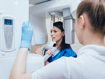
Common digital modalities include intraoral sensors for bitewing and periapical images and panoramic sensors for full-arch views. Intraoral sensors capture detailed views of individual teeth and surrounding bone while panoramic systems show broader anatomical relationships. Each modality has strengths that match different diagnostic tasks, from detecting cavities to assessing jaw structure.
Cone beam computed tomography (CBCT) provides three-dimensional imaging for complex cases such as implant planning and impacted teeth evaluation. CBCT exposes the patient to higher radiation than standard 2-D sensors but offers volumetric detail that can be critical for surgical planning. Clinicians select the modality that provides the information needed while taking into account diagnostic goals and radiation considerations.
A typical digital X-ray visit is brief: the clinician will position a small sensor in or near the mouth and ask you to remain still while the exposure is made. The process usually takes only a few seconds per image and images appear nearly instantly on the monitor. If an image needs adjustment, software tools can often clarify details without retaking the exposure.
There is no special preparation for most dental X-rays; you should follow any instructions about jewelry or removable appliances from your clinician. Lead aprons or thyroid collars may be used for additional protection based on the type of image and patient needs. Your dentist or hygienist will review the images with you and explain any findings and recommended next steps.
High-resolution digital images improve the detection of early decay, assessment of bone levels and evaluation of root anatomy and fractures. Software enhancements such as contrast adjustment and magnification help clinicians identify subtle changes that inform diagnosis. Consistently captured images also make it easier to track changes over time and evaluate treatment outcomes.
Digital radiography supports precise treatment planning for procedures like root canals, periodontal therapy and implant placement by revealing anatomical details. When combined with other records, annotated images help clinicians select approaches that reduce risk and increase predictability. Clear images also improve communication with specialists when coordinated care is needed.
Digital radiographs are stored as electronic files within the practice’s imaging system and integrated into the patient’s electronic dental record for easy retrieval. That structure allows clinicians to display prior images side-by-side with current ones to assess progression or healing. Standard file formats such as DICOM are often used to preserve image fidelity and metadata.
Secure electronic transfer makes it simple to share images with specialists, laboratories or other providers when referrals or consultations are required. Practices follow privacy protocols and secure channels to protect patient information during transmission. Reducing physical film handling also decreases the risk of lost or damaged records and speeds up collaborative care.
Although the radiation dose for digital radiography is low, no imaging modality is completely without risk and exposures should be justified by clinical need. Two-dimensional images can also have inherent limitations such as anatomical superimposition that may obscure certain findings. In some situations a three-dimensional CBCT scan or additional imaging techniques provide the missing perspective.
Technical factors such as patient movement or sensor positioning can produce artifacts that limit interpretation and sometimes require repeat imaging. High-quality results depend on proper technique and routine quality control of sensors and software. Clinicians balance the need for diagnostic detail with minimizing exposure by selecting the most appropriate modality and imaging protocol.
Digital radiography is particularly well suited to pediatric and routine monitoring because sensors require lower exposure and images are available immediately. Quicker imaging reduces time in the chair and lowers the chance of movement-related retakes, which is helpful with younger patients. Regular digital images enable clinicians to follow tooth eruption, monitor orthodontic progress and identify early changes in oral health.
For patients with ongoing restorative or periodontal treatment, digital records create a consistent baseline for comparison at future visits. This continuity helps detect slow changes that might benefit from earlier intervention. Clinicians can use the archived images to explain treatment needs and demonstrate progress to patients over time.
At Smiles N Beyond our team uses modern digital sensors and integrated imaging software to deliver diagnostic images that support efficient, patient-centered care. Those tools allow clinicians here in Wallington, NJ to review findings with patients in real time and make collaborative decisions during the visit. Using digital workflows also reduces manual handling and helps maintain consistent imaging protocols across appointments.
We combine technology with standard safety practices so imaging is performed responsibly and with attention to patient comfort. When advanced three-dimensional imaging is indicated, the team evaluates clinical benefit and selects the appropriate modality. Patients receive clear explanations of what the images show and how they influence recommended treatment.
Patients may request copies of their digital X-rays and many practices can export images in commonly used formats for transfer or personal records. Files such as DICOM preserve clinical detail and accompanying metadata while simpler formats like JPEG may be used for patient review. The practice can typically send images securely to a specialist or provide a copy for continuity of care.
To obtain records, contact Smiles N Beyond and specify the images or date range you need so the team can prepare a secure transfer. The staff will follow privacy and release procedures to ensure records are transmitted appropriately and in compliance with regulations. If you have questions about what will be shared or need help interpreting the images, the clinical team is available to review them with you.

Ready to schedule your next dental appointment or have questions about our services?
Getting in touch with Smiles N Beyond is simple. Our friendly team is here to help you schedule appointments, answer questions about treatment options, and address any concerns you may have. Whether you prefer to call our office, send an email, or complete our easy online contact form, we’re happy to assist you. Take the first step toward a healthier, more confident smile, contact us today and experience the difference personalized dental care can make.