Oral cancer can be aggressive, but one fact guides every modern approach to care: earlier detection saves lives. When abnormal changes in oral tissues are identified at an early stage, treatment options are broader and outcomes are significantly better. Regular screenings are a practical way to shift the odds in a patient’s favor, turning what might have been a late-stage diagnosis into a manageable condition discovered when intervention is most effective.
Risk factors such as tobacco and alcohol use, human papillomavirus (HPV), a history of previous oral lesions, or frequent sun exposure to the lips increase the need for careful monitoring. That said, oral cancer can occur in people without clear risk profiles, making routine exams an important safeguard for everyone. Awareness of symptoms — persistent soreness, unexplained lumps, or changes in color or texture of the mucosa — paired with dependable screening tools creates the best pathway to early action.
A screening is not a single moment of care but part of a broader preventive strategy. By combining patient history, visual inspection, palpation, and technology-assisted exams, clinicians can build a more complete picture of oral health. This layered approach reduces the chance that subtle, early-stage changes remain unnoticed until they progress into more advanced disease.
VELscope® is a handheld clinical device that enhances the clinician’s ability to spot tissue abnormalities that are hard to see under normal lighting. It uses a specific wavelength of blue light to cause healthy and abnormal oral tissues to fluoresce differently. That contrast often makes suspicious areas stand out during an in-office screening, so clinicians can mark them for closer observation, documentation, or follow-up testing.
It’s important to emphasize that VELscope® is designed as an adjunct — not a replacement — for a thorough oral exam. Visual inspection and palpation remain foundational, and VELscope® adds another layer of sensitivity. For many patients, the device reveals subtle changes in tissue architecture or metabolic activity that would otherwise be invisible during a routine check.
The benefits of this technology include a noninvasive way to expand diagnostic insight and a straightforward method for monitoring suspicious areas over time. Because the process is quick and painless, it fits seamlessly into hygiene visits and routine dental checkups, making advanced screening accessible without disrupting the flow of care.
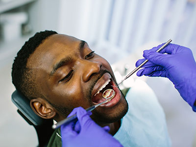
A VELscope® screening is fast and uncomplicated. After you’re comfortably seated, the clinician will perform a standard visual inspection and palpation to assess the soft tissues of your mouth, then dim the lights and use the VELscope® to scan the oral cavity. The blue light highlights tissue fluorescence patterns; healthy tissue typically appears with a characteristic glow, while areas that warrant attention may look darker or altered.
There is no special preparation required: no fasting, no anesthesia, and no recovery time. The exam itself takes only a few minutes and causes no discomfort. Clinicians often photograph or document findings so any changes can be compared over subsequent visits, which helps determine whether a suspicious area is stable, resolving, or progressing.
If a concerning area is identified, the clinician will discuss next steps clearly and calmly. That may include closer observation at more frequent intervals, referral to a specialist, or a biopsy to obtain tissue for definitive diagnosis. VELscope® helps clinicians identify where attention is needed; clinical judgment and follow-up testing determine diagnosis and treatment.
Interpreting VELscope® findings requires clinical experience and context. A dark or altered fluorescence pattern is a signal for further evaluation, not an automatic diagnosis. Tissue changes can result from a variety of benign causes — inflammation, trauma, or benign mucosal conditions — as well as premalignant or malignant processes. The clinician’s role is to separate routine, reversible changes from those that merit more urgent investigation.
Because the device increases sensitivity, it can also raise the frequency of observations that ultimately prove benign. That’s why follow-up protocols are essential. When the VELscope® highlights an area, dentists and hygienists will consider the patient’s medical history, risk factors, and any concurrent symptoms before recommending biopsy or specialist referral. This measured approach reduces unnecessary alarm while prioritizing patient safety.
When biopsy or referral is necessary, the goal is timely and accurate diagnosis. VELscope® shortens the path from suspicion to confirmation by clearly delineating which areas should be sampled or monitored, helping clinicians and patients make informed decisions without delay.
Including VELscope® in routine dental exams reflects a commitment to preventive, whole-person care. For patients, that means screening that goes beyond what the naked eye can reveal, combined with education about symptoms and behaviors that influence oral cancer risk. Clinicians can use the device to track changes across visits, create more informed treatment plans, and communicate more effectively about when intervention is necessary.
At Smiles N Beyond, this technology complements our patient-centered approach: it supports early detection while allowing clinicians to tailor follow-up based on individual risk. Whether a patient has identifiable risk factors or simply wants the reassurance of advanced screening, the addition of VELscope® permits more confident, evidence-informed decisions.
Ultimately, no single tool can eliminate risk, but using a technology like VELscope® alongside traditional examination techniques strengthens the overall screening process. Patients benefit from a higher degree of vigilance and a clearer plan for monitoring or action when tissue changes are identified.
In summary, VELscope® cancer screening adds a sensitive, noninvasive layer to routine oral examinations, helping clinicians detect and monitor tissue changes that might otherwise go unnoticed. It is an adjunctive tool that supports early detection, informed follow-up, and better patient outcomes when used within a comprehensive screening program. If you have concerns about oral cancer or would like to learn how this screening fits into preventive care, please contact us for more information.


VELscope cancer screening is an adjunctive diagnostic tool that helps clinicians identify abnormal oral tissues by using a specific wavelength of blue light. When the light is shone into the oral cavity, healthy and abnormal tissues fluoresce differently, making suspicious areas appear darker or altered compared with surrounding mucosa. That optical contrast can reveal subtle changes in tissue architecture or metabolism that are difficult to see with normal lighting alone.
The device is handheld and used in-office during a routine oral examination, typically after a standard visual inspection and palpation. Clinicians document fluorescence patterns and may photograph findings to compare across visits and monitor any changes over time. It is important to understand that VELscope is an adjunct and not a replacement for clinical judgment or biopsy when definitive diagnosis is required.
Early detection of oral cancer substantially improves treatment options and long-term outcomes because lesions found at an early stage are more likely to be localized and responsive to less invasive therapies. Detecting tissue changes before they progress allows clinicians to intervene sooner, reducing the risk of advanced disease and complications associated with later-stage treatment. Screening tools like VELscope contribute to earlier recognition by increasing the sensitivity of routine exams to subtle abnormalities.
Regular screenings are especially important because oral cancer can develop in people without classic risk factors, and some early lesions produce minimal or no symptoms. By combining patient history, visual inspection, palpation, and adjunctive technologies, clinicians create a layered strategy that decreases the chance of missing early-stage changes. This proactive approach supports timely follow-up and helps patients receive appropriate care when it is most effective.
Patients with known risk factors—such as tobacco or heavy alcohol use, a history of oral lesions, or significant sun exposure to the lips—should undergo regular oral cancer screening and closer monitoring. Human papillomavirus (HPV) exposure is also linked to oropharyngeal cancers, so patients with potential HPV risk factors may benefit from vigilant exams. Because oral cancer can appear without obvious risks, many clinicians recommend routine screenings for all adult dental patients as part of preventive care.
The appropriate screening interval depends on individual risk, clinical findings, and medical history; some patients may be screened at every hygiene visit while others follow a schedule based on documented observations. VELscope examinations are quick and nondisruptive, which allows clinicians to incorporate them into regular dental checkups when indicated. Discussing personal risk and recommended follow-up with your dental provider ensures screening frequency is tailored to your needs.
A VELscope screening begins with a standard oral examination that includes visual inspection and gentle palpation of soft tissues to assess any obvious abnormalities. The lights in the operatory are dimmed and the clinician uses the VELscope light to scan the oral cavity, observing fluorescence patterns across mucosal surfaces. Healthy tissues typically produce a characteristic fluorescence, while suspect areas may appear darker, discolored, or texturally different under the device's blue light.
There is no special preparation required, and the procedure is painless and completed within a few minutes, making it suitable to perform during routine visits. Clinicians often document findings with notes or photographs so that changes can be compared over time and decisions about monitoring or further testing can be evidence-based. If an area of concern is observed, the clinician will explain the significance, outline next steps, and recommend appropriate follow-up such as closer observation, specialist referral, or biopsy if indicated.
VELscope findings are not definitive diagnoses; rather, they highlight areas that warrant closer evaluation because the device increases sensitivity to tissue changes. Dark or altered fluorescence can result from a range of causes, including benign inflammation, trauma, or premalignant and malignant conditions, so context is essential. Clinical judgment, patient history, and additional testing determine whether an observed change represents a transient condition or a pathology requiring intervention.
When VELscope indicates a suspicious area, clinicians may schedule shorter-interval follow-ups to track stability or progression, refer to an oral medicine specialist, or arrange a biopsy for histologic confirmation. Prompt communication about findings and a clear, stepwise plan for evaluation help reduce unnecessary alarm while prioritizing timely diagnosis when needed. This layered approach ensures that VELscope guides decision-making without replacing the definitive information provided by tissue sampling and laboratory analysis.
Traditional visual inspection and palpation remain the foundation of oral cancer screening because they provide direct clinical context and tactile assessment of lesions. VELscope enhances rather than replaces those methods by revealing fluorescence patterns that may indicate subtle changes not visible under standard lighting. In many cases, the device increases the clinician's ability to localize areas for documentation and potential biopsy, improving the thoroughness of an exam.
Because VELscope increases sensitivity it can detect abnormalities earlier, but that heightened sensitivity also raises the likelihood of identifying benign changes that require interpretation and follow-up. A balanced screening strategy pairs the strengths of visual and tactile examination with adjunctive imaging to maximize early detection while minimizing unnecessary procedures. Clinicians trained in interpreting fluorescence patterns can integrate both approaches to form a more complete clinical picture.
VELscope screening is noninvasive and uses visible blue light, presenting no known radiation risk or lasting side effects for patients. The procedure is quick, causes no discomfort, and does not require anesthesia or recovery time, making it appropriate for most adult patients. Because exposure is brief and part of an in-office exam, it fits seamlessly into routine dental visits without special preparation.
Patients who have acute oral infections or significant sensitivity should communicate these concerns so the clinician can adapt the exam appropriately. Clinicians will combine VELscope results with the clinical exam and patient history to ensure safety and appropriate next steps. If you have specific health questions related to screening, your dental team can provide guidance tailored to your medical profile.
VELscope detects changes in tissue fluorescence and does not identify specific causes such as HPV; rather, it highlights areas with altered structure or metabolism that may result from a range of processes. Because HPV-associated oropharyngeal cancers often originate in deeper tissues of the throat, they can be more challenging to detect with an oral cavity fluorescence device alone. When clinicians suspect HPV-related disease based on location, symptoms, or patient history, they may use additional diagnostic pathways beyond VELscope to evaluate the concern.
A comprehensive evaluation for HPV-related cancer may include focused visual and endoscopic examination, imaging, and targeted biopsy when indicated. VELscope can still be a useful component of screening for visible oral mucosal changes and for guiding decisions about areas that need sampling. Discuss any HPV exposure history or throat-related symptoms with your clinician so they can select the most appropriate screening and diagnostic tools.
Interpreting VELscope results involves correlating fluorescence findings with clinical examination, medical history, and known risk factors to determine the level of concern. A stable or minor alteration may be observed and rechecked at defined intervals, while progressive or suspicious findings typically prompt referral or biopsy. Clear communication from the clinician about what was seen, why it matters, and how the practice will proceed helps patients understand the rationale behind recommendations.
Common next steps include increased surveillance, patient education about symptom awareness, referral to an oral medicine specialist, or obtaining a tissue biopsy for definitive histopathology. Timely scheduling and documentation support the ability to detect change over time, which is critical for distinguishing transient conditions from persistent or progressive lesions. Working together with your dental provider ensures that any recommended actions are appropriate, evidence-based, and aligned with your overall health status.
At Smiles N Beyond, we incorporate VELscope cancer screening into our preventive care protocol to enhance early detection efforts during routine dental visits. Our clinicians combine the device's fluorescence imaging with comprehensive visual exams, palpation, and review of medical history to create individualized monitoring plans. This integrated process helps our team in Wallington, NJ identify areas that require closer attention and communicate clear next steps to patients.
When a suspicious finding is identified, our approach emphasizes timely evaluation, careful documentation, and coordination with specialists when biopsy or advanced care is indicated. Providing education about symptoms and risk factors allows patients to participate actively in monitoring their oral health between visits. If you have questions about how VELscope screening fits into your care, our team is available to explain the process and determine the most appropriate screening schedule for you.

Ready to schedule your next dental appointment or have questions about our services?
Getting in touch with Smiles N Beyond is simple. Our friendly team is here to help you schedule appointments, answer questions about treatment options, and address any concerns you may have. Whether you prefer to call our office, send an email, or complete our easy online contact form, we’re happy to assist you. Take the first step toward a healthier, more confident smile, contact us today and experience the difference personalized dental care can make.